Living Architectures

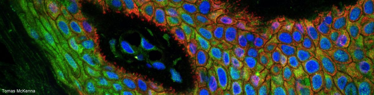
Amazing images of cells and pseudo cells on a miniature pattern replicating the facade of the Musée d’Orsay in Paris! The pattern was used to study the capacity of cells and cytoskeletal component to self-organise.

It was published by Manuel Théry and the CytoMorpho laboratory as a FocalPlane post and displayed as a night-show in Paris.

The conclusions of the experiments are highly inspiring, at the border between Biology, Art, Architecture and Philosophy. 😀

Conclusions

All of these experiments illustrate that it is by constantly breaking down that living systems are able to rebuild themselves by adapting to changes in their surroundings. The bonds between biological components are so fragile that they can be easily broken and rebuilt, which gives living structures great plasticity. This plasticity allows cells to constantly adapt to their environment. Unlike inert materials, in which the strength of the bonds allows them to resist and endure, it is the fragility of these bonds that allows living systems to constantly reinvent themselves. But their plasticity is also their weakness. If the balance between destruction and construction is not perfect, the system cannot hold together and dies quickly.

Cells thus use their internal dynamics to explore every possibility, without goal or intention, simply because it is possible. Anything too unstable will collapse and make way for other attempts. Cells therefore spend their time playing with the limits of their stability. Most of them will die as a result. We are today the product of an evolution paved by the death of trillions upon trillions of cells. These adventurers did not attempt to achieve a specific goal that would have made them more robust or more efficient. They tried everything made possible by their internal dynamics.

Image: Nevena Morel


Train-the-Trainer course 2026

Do you train others to use light microscopy and would like to improve your training?

Together with Rhonda R. Powell from Clemson University in the USA, I will give the second edition of the online course “Magnify your impact: Train the Microscopy Trainer”.

This practical, interactive, online course is intended for anyone with experience training people to use light microscopes, including Core Facility Staff and Industry Applications Specialists.

During the course, you will focus on improving your OWN training. By the end of the course, you will be able to:

  • Write a list of learning objectives for your own microscopy training
  • Incorporate new pedagogical tools in your microscopy training
  • Evaluate the quality of your training using assessments and feedback

The application page is open until the 1st of August.

SeeDB-Live: Clearing live samples with BSA

Amazing talk at the ELMI in Coimbra!

Takeshi Imai presented SeeDB-Live, a new clearing technique for live samples. The refractive index is 1.363 and the technique works exceptionally well in loose tissues like brain but allows also 2 times deeper imaging in live organoids. They could even image calcium with multiphoton repeatedly over several months in brain of live mice!

The paper was just published in Nature.

FLASH-PAINT: nm resolution, fast and highly multiplexed!

Just heard this amazing Eurobioimaging talk by Florian Shueder from EFPL. They published FLASH-PAINT, an improved version of DNA-Paint.

FLASH-PAINT combined with fluorogenic probes, eraser oligos and EXCHANGE-PAINT has many advantages over DNA-PAINT:

    • It keeps the very high resolution of just a few nm.
    • It is very fast.
    • It enables imaging with a widefield system instead of in TIRF.
    • It enables high multiplexing (they demonstrate 12 rounds) without needing to wash.

They demonstrate 12-plex imaging with 25nm resolution in just 2h!


Reproducibility?

Have you ever heard of the Antibody reproducibility crisis?

Well here is a new chapter in this sad saga and this one is about the possible image manipulation of the Western Blots on the datasheets of commercial antibodies.

There is a strong need for clearer ethical guidelines and more education.

Free, online microscopy course by the LCI core facility

Dear all

Kindly forward to anyone who might be interested.

It is my pleasure to invite you to follow our free (no registration) online microscopy course.

The course schedule and Zoom link are found our course webpage.

The public program (everything in blue in the course schedule) starts next week on Thursday 22th of Jan and runs until the 13th of February.

Here is a selection of what we will talk about:

  • Optics and image formation,
  • How microscopes work
  • Objectives and refraction index
  • Cameras and detectors, Noise and background, Bit depth and saturation
  • Sample preparation, Immunostaining, Clearing
  • Resolution and contrast, Nyquist sampling, how to choose microscope settings
  • Data handling, how to make article figures, Requirements for image analysis, Colocalization, Ethics
  • Image processing and analysis

All lectures will be broadcasted live on Zoom, recorded then posted the same day on our Youtube channel.

The course syllabus with the Learning Objectives can be downloaded here.

Do not miss the kind testimonies on our course webpage! 🙂

The LCI core facility team, Stockholm, Sweden

Jobs opening at QUAREP-LIMI

A rare opportunity for those interested in contributing to Quality and Reproducibility within Light Microscopy (Quarep-LiMi consortium).

The QUAREP-LiMi network is currently recruiting for three positions related to Light Microscopy Metadata, Quality Control, and Community Management to work within the QUAREP-LiMi initiative (quarep.org):

    • Lead Specialist – Light Microscopy Model (LiMi-Model)
    • Lead Specialist – Validation Team
    • Community Coordinator – QUAREP-LiMi

You can find more details about the positions here.

Please send your application, including supporting documents (CV, motivation letter, references, Certificates), and anything else that might help our decision, by 15 January 2026 at the latest, using this application form.

3D imaging symposium

Dear all

An interesting whole-day 3D imaging symposium at Scilife on the 4th of December. Many really inspiring talks! 🙂

Time to apply to the LCI Microscopy course! :)

Dear all

You are now welcome to apply to join the LCI light microscopy course (21 Jan – 13 Feb 2026) ‘Microscopy: improve your imaging skills – from sample preparation to image analysis’ (6 credits).

The purpose of this fully online yet very applied course is to provide PhD students and researchers with enough theoretical and practical knowledge about their OWN sample and their OWN microscope, to enable them to:

    1. Prepare their sample and formulate their scientific question in a way that is suitable for reliable data extraction.
    2. Properly use the software and hardware available on the microscope they use in their lab/facility.
    3. Design their experiment based on image analysis requirements.

Application deadline 5th of November.

Course webpage.

Course syllabus, description and registration. Please carefully check the eligibility criteria and make sure to include all required items in your application.

Those who cannot register to the course can anyway follow any of the lectures (in blue on the schedule) as they will be publicly broadcasted live on Zoom and accessible to anyone without registration. The schedule and zoom link are on the course page.

Kindly help us spread the word. 🙂

The LCI team

Online course for microscopy facility staff: “Magnify Your Impact: Train the Microscopy Trainer.”

Dr. Sylvie Le Guyader (sylvie.le.guyader@ki.se) and Ms. Rhonda Powell (rhondar@clemson.edu) invite you to apply to the online course: “Magnify Your Impact: Train the Microscopy Trainer.”

This practical, interactive, online course is intended for anyone with experience training people to use light microscopes, including Core Facility Staff and Industry Applications Specialists.

During the course, you will focus on improving your OWN training. By the end of the course, you will be able to:

  • Write a list of learning objectives for your own microscopy training
  • Incorporate new pedagogical tools in your microscopy training
  • Evaluate the quality of your training using assessments and feedback

Please see the attached flyer for details and a link to the application, or visit https://tinyurl.com/magnifyyourimpactapplication

The application deadline is September 20, 2025. Applications will be reviewed in the order they are received. Questions about the application can be directed to Rhonda.

The course is sponsored by BNMI and Nordforsk.

Follow us! :-)

Type your email address below to subscribe to our blog!